Figure 2. Membrane integrity is not compromised by PlyC, and internalization is mediated by the PlyCB subunit.
(a) A549 epithelial cells were permeabilized with 0.02% Triton X-100 (left panel), as shown by staining with propidium iodide (red) and DAPI (nucleus, blue), whereas cells pre-incubated with 100 μg/ml PlyC for 1 hr showed no damage (right). Scale bar is 5 μm. (b) Fluorescently labeled PlyC (left panel) and PlyCB (middle panel), but not PlyCA (right panel), are internalized in A549 cells upon incubation, as shown with protein (5 µg/ml) conjugated with AlexaFluor555 (green stain). Cells were incubated in serum-free F12K medium for 30 min at 37°C, fixed with 4% PFA in PBS and subsequently stained with DAPI (blue). Arrows indicate internalized PlyC or PlyCB in vesicle-like structures with an average size of 0.5 μm. Scale bar is 10 μm. (c) Confocal microscopy of internalized PlyC colocalized with intracellular Spy. Nucleus (blue, DAPI), Spy bacteria (green, AlexFluor 488 conjugated wheat germ agglutinin), PlyC (red, AlexFluor 555 conjugated), and actin filament (magenta, AlexFluor 647 conjugated phalloidin) are shown in the same focal plane. PlyC (red) is colocalized with Spy (green) in the merged image. The maximum intensity projection shows all Z-stacks simultaneously. Scale bar, 10 μm.
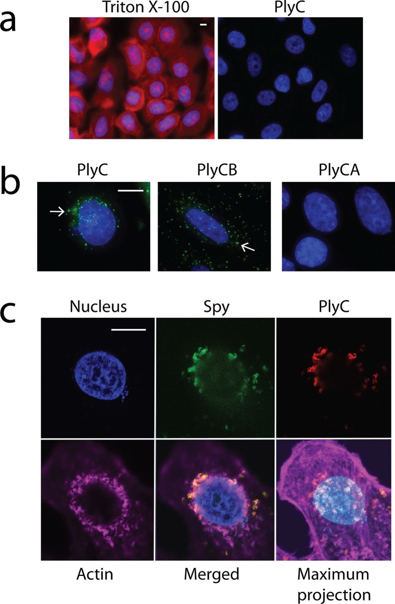
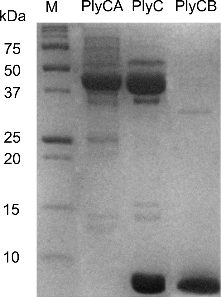
Figure 2—figure supplement 1. Purification of PlyC and its individual subunits.