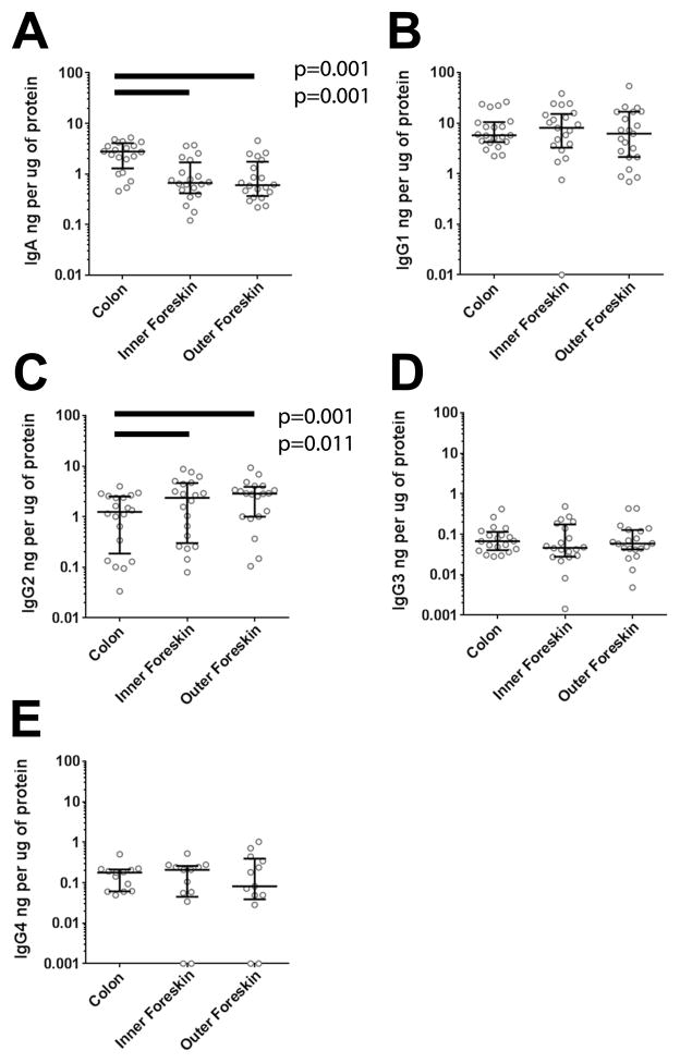
Figure 1. The foreskin contains more IgG2 and less IgA than colonic mucosa.
Twenty paired foreskin and colonic tissue samples were homogenized and assayed by multiplex bead array for A) IgA, B) IgG1, C) IgG2, D) IgG3, and E) IgG4. All Ig concentrations were normalized to μg of protein in the tissue biopsy. Bars indicate median with IQR for each tissue. Lysate buffer designed for homogenization of skin samples prevented detection of IgM and IgE standards, and thus their quantitation was not included in the figure. Only significant differences in Wilcoxon post-test after Bonferroni correction for three comparisons are reported in the figure.

