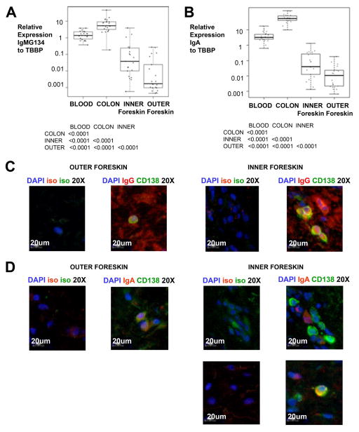
Figure 4. Foreskin tissue contains antibody secreting cells.
A–B) Real-time PCR comparing 20 paired colonic biopsies, blood, and inner and outer foreskin was used to quantitate mRNA transcription with a primer that amplifies A) IgM, IgG1, IgG3, and IgG4 (IgMG134); and B) IgA; normalized to TATA-box binding protein (TBBP). Only significant differences in Wilcoxon post-test after Bonferroni correction for six comparisons are reported beneath each graph. Individual measurements are represented by circles. Box plots show the median and IQR, with whiskers extending to the range of the data for each tissue. C) Representative 20× magnification immunofluorescence microscopy images of paired inner and outer foreskin sections stained with DAPI nuclear counterstain (pseudocolored blue), CD138 (pseudocolored green) and IgG or their isotype control (both pseudocolored red). D) Selected 20× magnification immunofluorescence microscopy images of paired inner and outer foreskin sections stained with DAPI nuclear counterstain (pseudocolored blue), CD138 (pseudocolored green) and IgA or their isotype control (pseudocolored red). In C) and D), we analyzed ~2.6mm2 sections from 18 individuals, but photos represent only 1/200 of the area used to quantitate ASCs.

