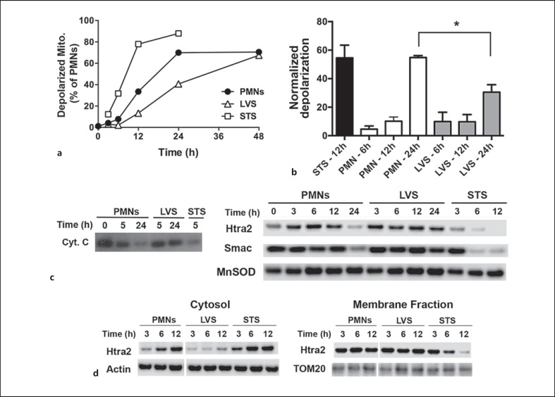
Fig. 2.
F. tularensis sustains neutrophil mitochondrial integrity. PMNs were left untreated, infected with LVS or treated with STS as indicated. a, b Loss of mitochondrial membrane potential was quantified by JC-1 staining. a A representative time course of mitochondrial depolarization. Mito = mitochondria. b Pooled data normalized to the fluorescence at time zero, showing the mean ± SEM of 3 independent experiments. * p < 0.05. c, d Outer mitochondrial membrane permeabilization was assessed by subcellular fractionation. c Immunoblots of cytochrome c (Cyt. C), Htra2 and Smac retained in mitochondria-containing fractions at each time point and are representative of at least 4 independent experiments. d Matched immunoblots of cytosol and mitochondria-containing membrane fractions show delayed release of Htra2 into the cytosol during LVS infection and are representative of at least 4 independent experiments. β-actin, MnSOD and TOM20 were used as loading controls.

