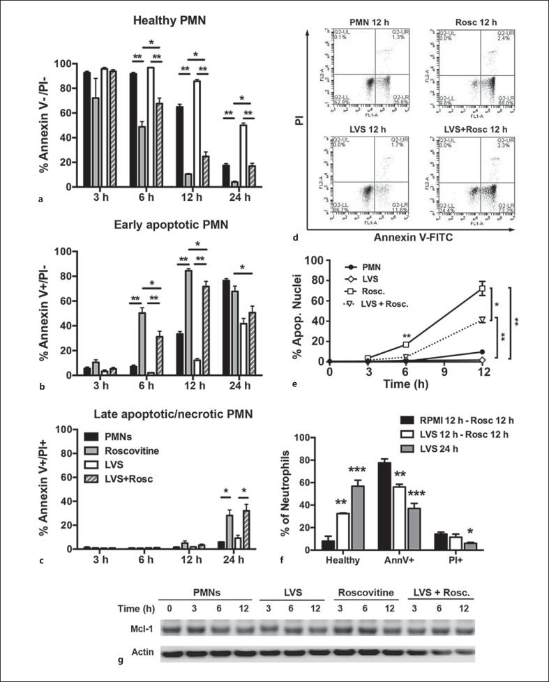
Fig. 6.
F. tularensis attenuates the ability of R-roscovitine to induce neutrophil apoptosis. Effects of R-roscovitine on apoptosis of control and LVS-infected PMNs were quantified by Annexin V-FITC/PI staining or by analysis of nuclear morphology. a-e, g R-roscovitine was added 1 h after LVS infection. a-c Graphs show the percentage of PMNs at each time point and condition that are healthy (Annexin V-/PI-), in early apoptosis (Annexin V+/PI-), or have progressed to late apoptosis/secondary necrosis (Annexin V+/PI+), respectively. Data are the mean ± SD of 5 independent experiments. ** p < 0.001; * p < 0.05. d Representative flow cytometry plots at the 12-hour time point. e Percentage of PMNs with an apoptotic nuclear morphology. Data are the mean ± SD of 3 independent experiments. * p < 0.05; ** p < 0.01. f PMNs were left in medium or infected with LVS for 12 h prior to treatment with R-roscovitine for 12 h, or were infected with LVS for 24 h in the absence of R-roscovitine. Annexin V-FITC/PI staining results are the mean ± SD of 3 independent experiments. ** p < 0.01 for LVS at 12 h - R-roscovitine at 12 h versus both other treatments. *** p < 0.001; * p < 0.05 for LVS at 24 h vs. RPMI at 12 h - R-roscovitine at 12 h. g Immunoblots of PMN lysates prepared at the indicated time points were probed to detect Mcl-1 with β-actin as the loading control. Data are from 1 experiment representative of 3 independent determinations. Rosc. = R-roscovitine.

