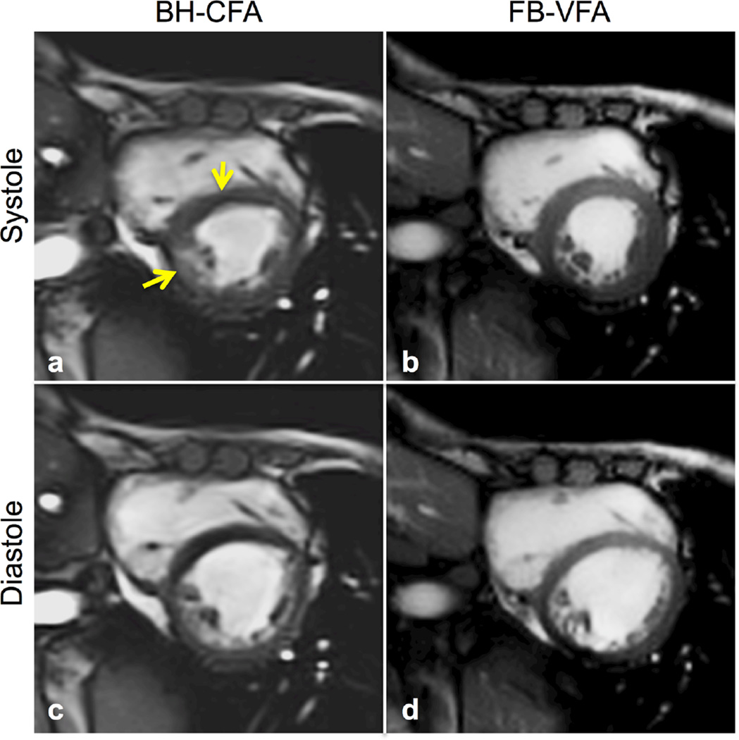
FIG. 3.
Systolic (a,b) and diastolic (c,d) cardiac images from a patient with DMD acquired during instructions for breath-holding with constant flip angle (BH-CFA) acquisition (a,c) compared with free-breathing, variable flip angle (FB-VFA) cardiac cine images (b,d). The myocardial septum has notable image artifacts related to the patient’s poor breath-hold abilities (yellow arrows) that are not observable in the FB-VFA images.

