Fig. 8.

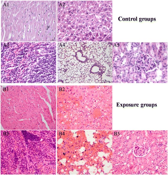
The histopathology (×400) of tissues after exposure CdS to mice (A1–A5 and B1–B5 for the heart, liver, spleen, lung, and kidney, respectively)

The histopathology (×400) of tissues after exposure CdS to mice (A1–A5 and B1–B5 for the heart, liver, spleen, lung, and kidney, respectively)