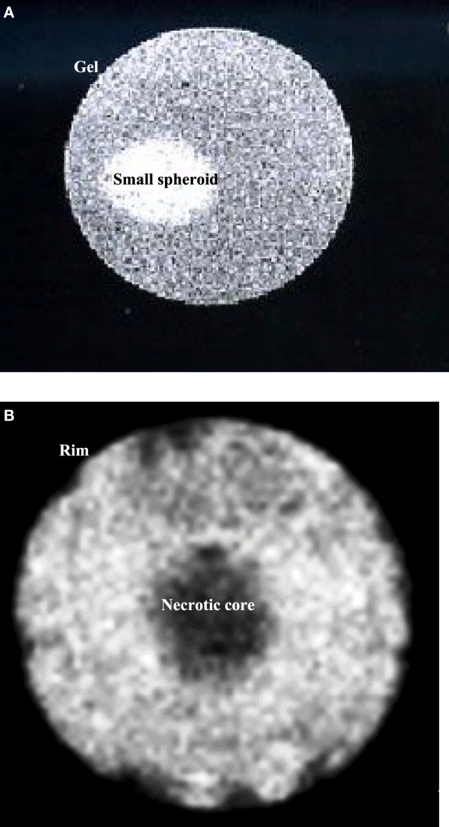
Figure 1.

(A) Sagittal image of a small spheroid with a diameter of about 0.5 mm, inserted in agarose gel. (B) Sagittal image of a spheroid after 10 days in culture. The diameter is about 1.8 mm. It is possible to identify two different regions: a necrotic core and a viable rim. Both images were acquired with an MSME sequence.
