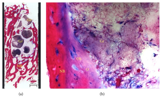
Figure 9.
Histological examination. Saw cut (a) of the core biopsy showing formation of new bone (NB). Graft particles (Gr) are embedded in connective tissue and bone trabeculae (Azur II and Pararosanilin, original magnification ×50); (b) osteoblasts (Ob) forming osteoid (O) and adding new bone (NB) to the surface of partially disintegrated graft particles (Gr) (Azur II and Pararosanilin, original magnification ×200).

