Figure 2.
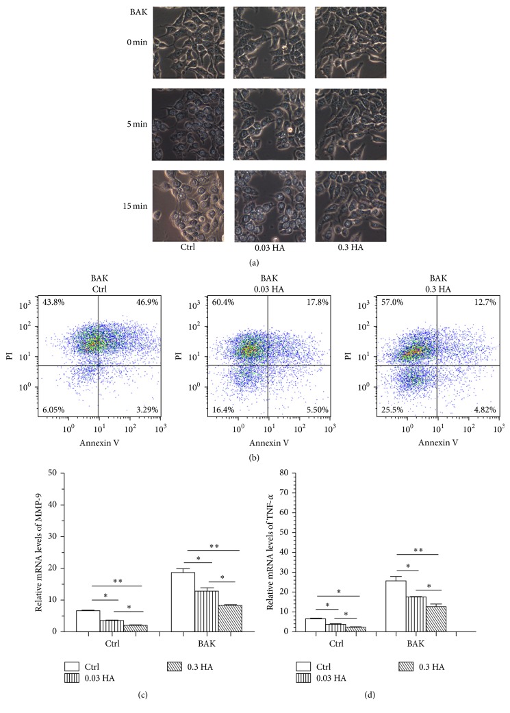
Hyaluronate decreased BAK-induced cell apoptosis and the expression of inflammatory cytokines. Microscopic images (a) of the changes in cell morphology in response to the hyperosmolar conditions were examined in the control, 0.03 HA, and 0.3 HA groups. Flow cytometry (b) was performed to analyse cell apoptosis, and real-time PCR (c) and (d) were used to compare the expression levels of MMP-9 and TNF-α among the three groups. Magnification: ×100. ∗ p < 0.05; ∗∗ p < 0.01; ∗∗∗ p < 0.001. The data are the mean ± SEM and represent individual experiments, each including 5 samples/group/time.

