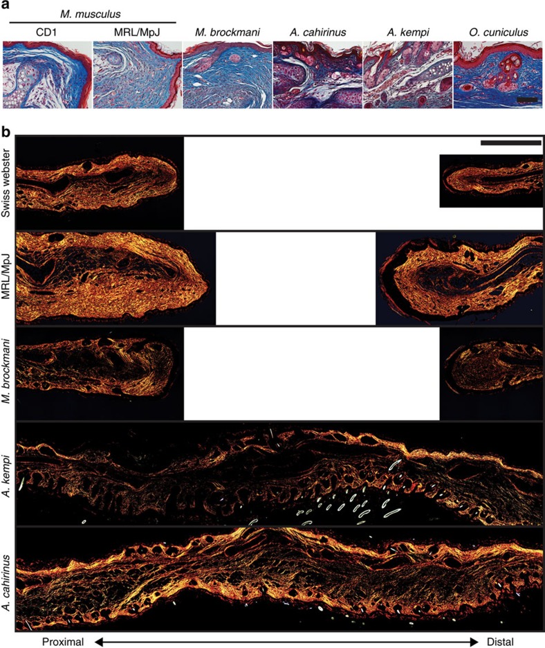
Figure 3. Regenerating species restore dermal architecture whereas non-regenerating species form scar tissue in the dermis.
(a) Representative images showing the dermis at D85 (D96 for MRL/MpJ) stained with Masson's trichrome. Regenerating species (for example, A. cahirinus, A. kempi and O. cuniculus) regenerate full-thickness skin, including bilayer dermis and epidermis with hair follicles and sebaceous glands. Non-regenerators (for example, M. musculus strains—CD1 and MRL/MpJ, and M. brockmani) form scar tissue beneath the epidermis evident as dense, parallel bands of collagen. In Mus strains, no new hair follicles or sebaceous glands are evident distal to the wound. In contrast, new hair follicles and sebaceous glands are present in M. brockmani. (b) Representative images of D85 tissue stained with picrosirius red viewed under polarized light to visualize thick (red) and thin (green) collagen fibres. In non-regenerating species the thick, parallel fibres with very little observed thin fibres were indicative of a scar. However, in regenerating species, collagen architecture is more representative of uninjured collagen pattern of a thick and thin network of uninjured tissue. All images are representative of at least n=3 biological replicates per species. Scale bars, 50 (a) and 500 μM (b).

