
Figure 1. Imaging of APC through polarized light microscopy and UHR FE-SEM.
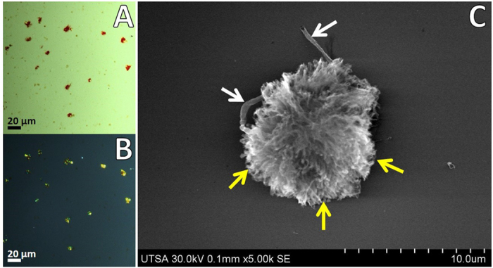
APC were stained with Congo red and analyzed under polarized light for characteristic green birefringence of amyloid protein aggregates. (A) Transmitted light. (B) Polarized light. (C) High resolution imaging of APC by FE-SEM revealed the details of compact aggregates with fibrillar radial projections (yellow arrows) and presence of blood vessels (white arrows).