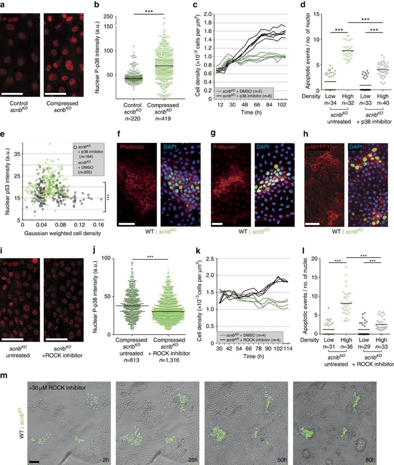
Figure 4. During mechanical cell competition ROCK activates p38 leading to p53 elevation.
(a) Phosphorylated p38 (P-p38) staining in pure scribKD cells +/− compression. (b) Single-cell nuclear P-p38 intensity from images as in a; black bars=median. (c) Time-resolved density measurement of growing scribKD cells +/− p38 inhibitor. (d) Quantification of cell death (cleaved Caspase-3) of scribKD cells with or without compression and +/− p38 inhibitor; black bars=mean; pooled data from three biological replicates across two independent experiments. (e) Single-cell nuclear p53 signal intensity in competing scribKD cells +/− p38 inhibitor, plotted against cell density. (f–h) F-Actin (phalloidin-stained, f), phosphorylated Myosin-II (P-Myosin, g) and phosphorylated MYPT1 (p-MYPT1, h) are elevated in compacted GFP-labelled scribKD cells compared with wild-type (WT) cells during competition. (i) P-p38 staining in compressed scribKD cells +/− ROCK inhibitor. (j) Single-cell nuclear P-p38 intensity from images as in i; black bars=median. (k) Time-resolved density measurement of growing scribKD cells +/− ROCK inhibitor; two independent repeats. (l) Quantification of cell death (cleaved Caspase-3) in scribKD cells with or without compression and +/− ROCK inhibitor; black bars=mean; pooled data from three biological replicates across two independent experiments. (m) Stills from time-lapse movies of WT and GFP-labelled scribKD co-cultures treated with ROCK inhibitor (30 μM), see corresponding Supplementary Movie 12; n=cell number in b,e and j or n=number of imaged fields of cells in c,d,k and l. ***P<0.0005 by KS test.

