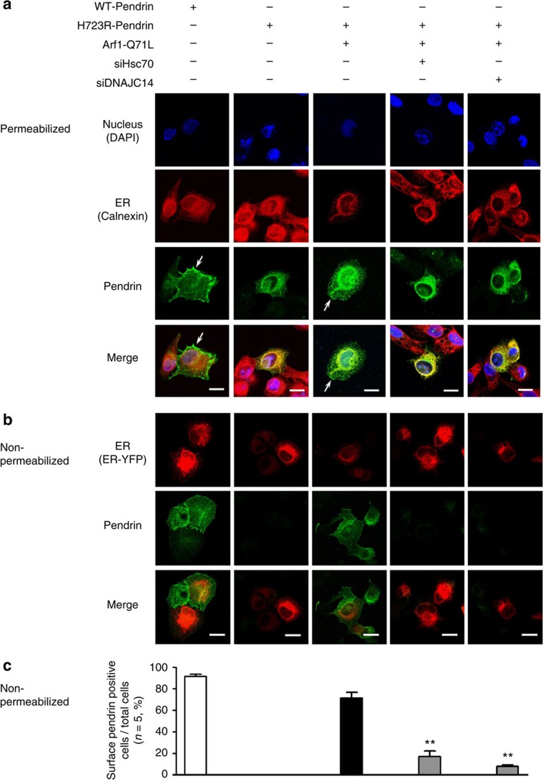
Figure 6. Knockdown of Hsc70 and DNAJ14 inhibits the cell-surface expression H723R-pendrin induced by Arf1-Q71L.
(a) Cellular localization of pendrin was examined in PANC-1 cells permeabilized with ethanol and acetone. Cells stained for WT- and H723R-pendrin were co-stained for the ER marker protein calnexin. Arrows indicate the cell-surface expression of WT- or H723R-pendrin. Nuclei were counterstained with 4′,6-diamidino-2-phenylindole (DAPI). Scale bar, 20 μm. (b,c) Cell-surface expression of WT- and H723R-pendrin was examined in non-permeabilized PANC-1 cells (4% formaldehyde fixation) using antibodies against the extracellular N terminus of pendrin8 (Abcam, ab66702). The plasmid encoding the ER marker protein ER-yellow fluorescent protein (YFP) was co-transfected. Arf1-Q71L induced the cell-surface expression of H723R-pendrin, which was blocked by siRNAs targeting Hsc70 and DNAJC14. Quantification of the ratio of cells expressing pendrin on the cell surface relative to total cells in five independent experiments is shown in c. Scale bar, 20 μm. **P<0.01 by one-way analysis of variance, difference from H723R-Pendrin+Arf1-Q71L alone (lane 3), n=5.

