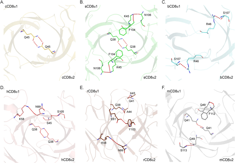
Figure 2. The inter-chain hydrogen bonds between the monomers of the CD8αα dimers.
The CD8αα structures and colours are the same as above. The hydrogen bonds are shown as red dashed lines. (A–F) Hydrogen bonds in chicken, swine, bovine, human, monkey and mouse CD8αα homodimers. The residues forming these hydrogen bonds are shown in stick models. The numbers and locations of hydrogen bonds in the six CD8αα homodimers are not conserved.

