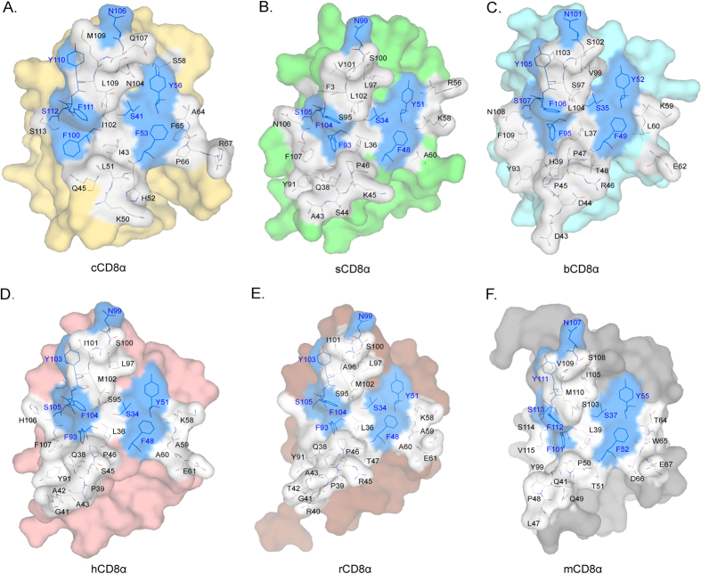
Figure 3. The residues in the interfaces of the six known CD8αα structures.
Residues in the interfaces of the six known CD8αα homodimers are shown as stick models. The conserved residues are coloured blue, and non-conserved residues are coloured white. (A–F) The interfaces and residues composing chicken, swine, bovine, human, monkey and mouse CD8αα homodimers.

