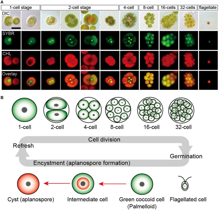
Figure 2.
Life cycle of H. pluvialis. (A) Fluorescence microscopy images, showing the 1- to 32-cell stages, and the flagellated stage. DIC, differential interference contrast image; SYBR, SYBR Green I-stained cells (green); CHL, chlorophyll autofluorescence (red); and Overlay, overlaid images of SYBR and CHL. (B) Illustration of life cycle of H. pluvialis. Refresh, when old cultures are transplanted into fresh medium, coccoid cells undergo cell division to form flagellated cells within the mother cell wall. Germination, Flagellated cells settle and become coccoid cells. Continuous and/or strong light accelerate the accumulation of astaxanthin during encystment (red arrows). Figure reproduced from Wayama et al. (2013) distributed under the terms of the Creative Commons Attribution License.

