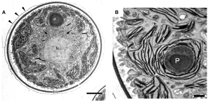
Figure 4.
Transmission electron micrographs of green vegetative cells of H. pluvialis. (A) General ultrastructure. The cell wall is surrounded by extracellular matrix (arrowheads). Arrows indicate astaxanthin granules. (B) Chloroplast and pyrenoid. C, chloroplast; CW, cell wall; N, nucleus; P, pyrenoid. Scale bars in (A,B): 5 and 1 μm, respectively. Figure reproduced from Wayama et al. (2013) distributed under the terms of the Creative Commons Attribution License.

