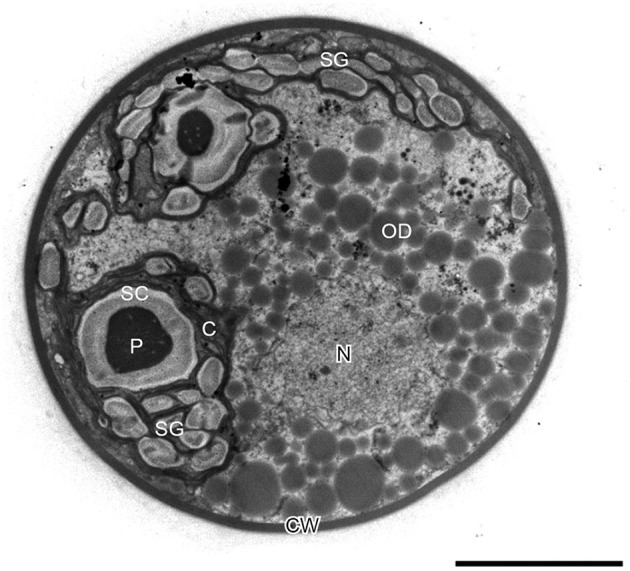
Figure 5.

Transmission electron micrograph of intermediate H. pluvialis cell (general ultrastructure). C, chloroplast; CW, cell wall; N, nucleus; OD; oil droplet; P, pyrenoid; SC, starch capsule; SG, starch grain. Scale bar: 5 μm. Figure reproduced from Wayama et al. (2013) distributed under the terms of the Creative Commons Attribution License.
