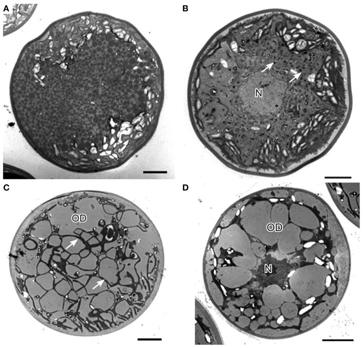
Figure 6.
Transmission electron micrographs of H. pluvialis cyst cells. (A) General ultrastructure of cyst cells, showing small granules that contain astaxanthin. (B) General ultrastructure of a cyst cell, showing astaxanthin accumulation in oil droplets. (C) General ultrastructure of a cyst cell, showing large oil droplets. Chloroplasts localize in the interspace between oil droplets (arrows). (D) Some oil droplets are fused. C, chloroplast; N, nucleus; OD, oil droplet. Scale bars in (A–D): 5 μm. Figure reproduced from Wayama et al. (2013) distributed under the terms of the Creative Commons Attribution License.

