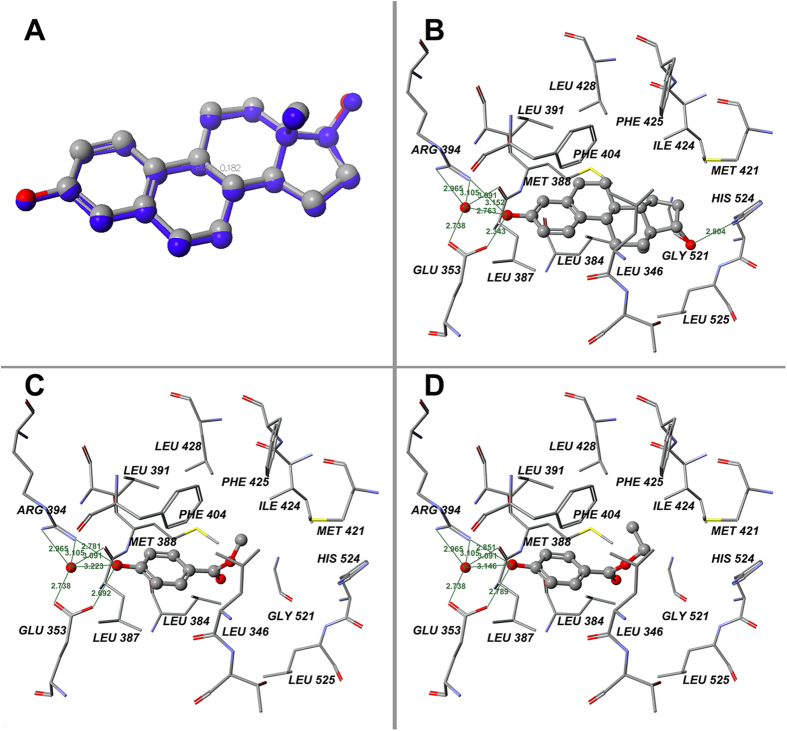
Figure 4. Results of docking calculations on hERα-LBD.
(A) Validation of docking of 1ERE with E2: docked ligand (blue) and ligand of crystal structure at their absolute positions in binding pocket; (B) Binding positions of original ligand E2 on hERα-LBD in x-ray structure (1ERE chain A) template; (C) Binding positions of MP; (D) Binding positions of EP. Green lines indicate hydrogen bonds between chemicals and amino acid residues. Green numerals indicate distances between two atoms (Å).

