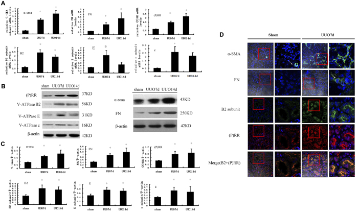
Figure 1. Expression of (P) RR, Vacuolar ATPase (V-ATPase) B2, E, c subunits, and α- SMA, fibronectin (FN) in kidney of SD rats at day 7 or 14 after UUO and SD rats with sham-operation.
(A) Real-time PCR analysis showed mRNA expression of α-SMA, FN, (P) RR and Vacuolar ATPase (V-ATPase) B2, E, c subunits. Values are means ± SD; n = 4. *P < 0.05 vs. the sham group. (B) Western blot analysis of protein expression of (P) RR, V-ATPase subunits (B2, E, and c) and α- SMA, FN, showed higher expression of (P) RR, V-ATPase B2, E, c subunits, and α-SMA, FN in UUO rat. (C) Graphic representation of the protein levels of α-SMA, FN, (P) RR, V-ATPase B2, E, and c subunits at SD rats at day 7 or 14 after UUO and SD rats with sham-operation. Values are means ± SD; n = 4. *P < 0.05 vs. sham group. (D) immunofluorescence showed upregulation of (P) RR, V-ATPase B2, α-SMA and FN at SD rats kidney at day 7 after UUO respectively (×200), and the higher-magnification confocal microscopy images of the contents in red box (showed at right panel of Sham or UUO, ×400) showed that the B2 subunits and (P) RR significantly increased in apical region of tubular cells after UUO, and marked colocalization after UUO. Nucleus was stained with 4, 6-diamidino-2-phenylindole (blue). Confocal ×40 ocular.

