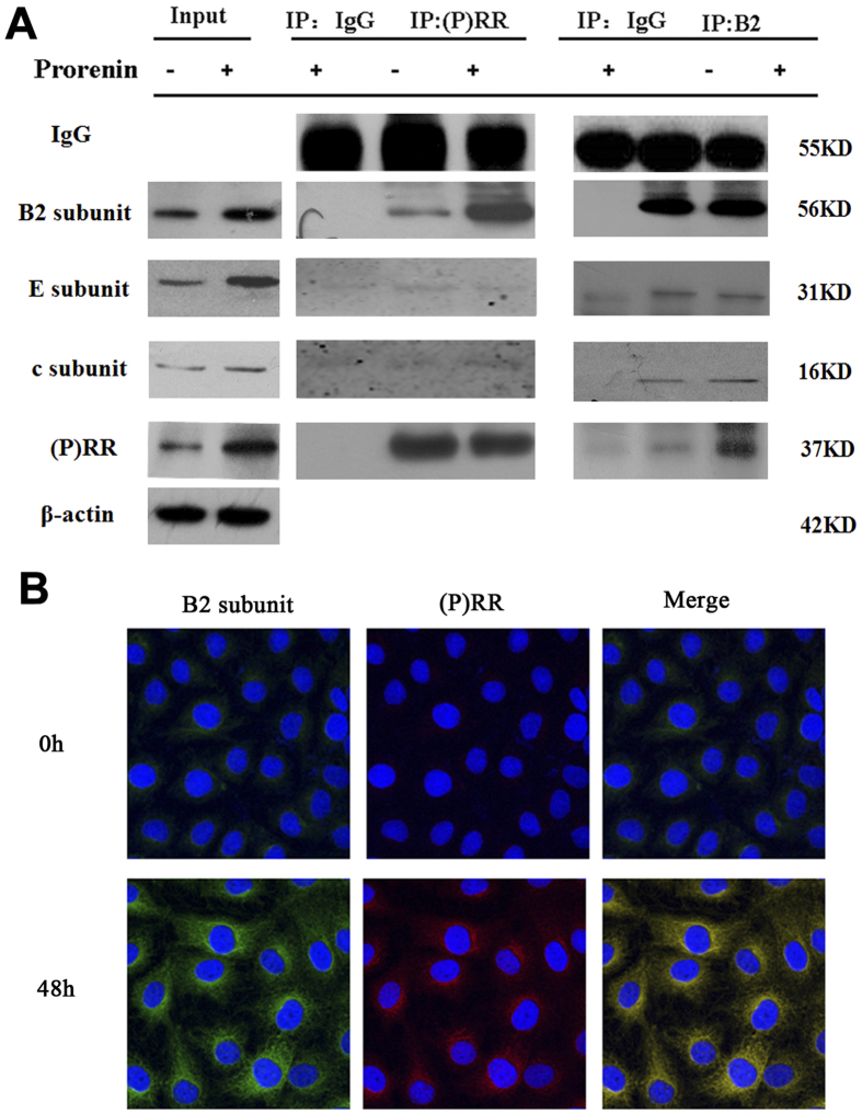
Figure 6. Interaction between (P) RR and V-ATPase B2 by co-immunoprecipitates and co-immunostaining.
(A) 800 μg of whole cell homogenates from NRK-52E cells stimulated by prorenin (100 pmol/L, 0 and 48 h) were prepared with 4 μg antibodies or 2 μg IgG as a negative control. Western blots demonstrating the presence of the V-ATPase B2 subunit (56KD) in anti-(P) RR (IP: (P) RR, Rabbit polyclonal antibody) immunoprecipitates, and this effect was upregulated after prorenin stimulation (100 pmo/L, 48 h); meanwhile, the presence of the (P) RR (37KD) and other subunits of V-ATPase (E subunits, 31KD; c subunits, 16 kD) were measured by Western blots after immunoprecipitated by anti-V-ATPase B2 subunit (IP: B2, mouse monoclonal antibody), and this effect was also upregulated after prorenin stimulation (100 pmo/L, 48 h). (B) Co-immunofluorescence results showed marked colocalization of (P) RR (red) and vacuolar ATPase B2 subunit (green) in NRK52E cells after prorenin stimulation. Nucleus was stained with 4,6-diamidino-2-phenylindole (blue). Confocal ×40 ocular.

