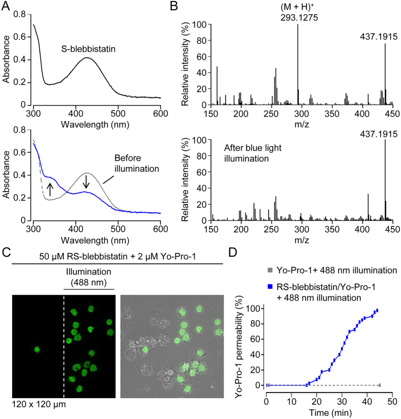
Figure 4. Photodegradation and phototoxicity of S-blebbistatin.
(A) Absorption scans of S-blebbistatin (50 μM in a 1:1 mixture of DMSO and PBS) before and after 2 h illumination with blue light (450–490 nm). In the lower plot, the absorption scans before (gray; dashed line) and after (blue line) blue light illumination of S-blebbistatin are superimposed. Accordingly, the arrows indicate the change in absorption induced by blue light illumination. (B) Mass spectra before and after blue light illumination. The calculated mass of the parent molecule S-blebbistatin ionized by protonation (designated (M + H)+) is 293.1275. The m/z ratio of 437.1915 probably corresponds to 2(M - phenyl group) with loss of a methyl group and ionization by Na+. (C) Fluorescence image (on the left) showing Yo-Pro-1 staining of nuclei, an indicator of loss of plasma membrane integrity, after 45 min intermittent illumination with blue (488 nm) light. The peripheral human mononuclear cells, enriched for monocytes by excessive washing (to remove nonadherent cells), were scanned by serial z-stacks (1 stack = 20 optical slices) obtained at a rate of 4 stacks/min. On the right is an overlay of fluorescence and brightfield images. Note that, after 45 min illumination, the microscope stage was moved to the right to reveal cells (left of the vertical dashed white line) which had not been illuminated. (D) Cummulative plot of Yo-Pro-1 permeability in individual cells. In the presence of RS-blebbistatin (solid blue line and dots), the first cell became Yo-Pro-1 positive after 17 min illumination. In the absence of blebbistatin (dashed grey line and dots), blue (488 nm) light illumination did not induce Yo-Pro-1 permeability.

