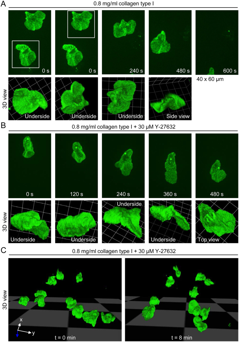
Figure 6. 3D morphology of human monocytes migrating in a loose collagen type I matrix.
(A) Projected (extended focus) fluorescence images (40 × 60 μm; upper panel) of control monocytes migrating in a loose collagen type I matrix (0.8 mg/ml collagen), obtained by time-lapse spinning disk confocal microscopy, and corresponding reconstruction of 3D morphology (lower panel). (B) Projected (extended focus) fluorescence images (40 × 60 μm; upper panel) of a monocyte migrating in the presence of the ROCK inhibitor Y-27632, and corresponding reconstruction of 3D morphology (lower panel). (C) 3D morphologies of monocytes in the presence of Y-27632 at two time points.

