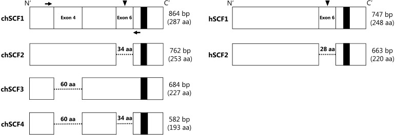
Fig. 1.
Isoforms of chSCF. Comparison of protein structures of chSCF1–4 and the secreted and membrane-bound forms of human SCF (hSCF1 and hSCF2, respectively). Dashed lines indicated deleted regions, and filled squares in the C’ region indicate the transmembrane domain. Arrowheads and black arrows indicate the putative proteolytic cleavage site and the forward and reverse primers used for Fig. 2B, respectively.

