Fig. 4.
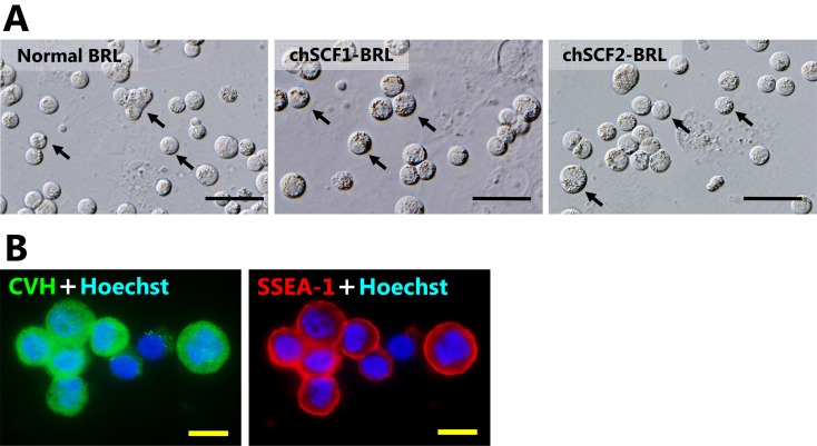
Characterization of cultured PGCs. A) Morphological characteristics of PGCs cultured on chSCF1-BRL, chSCF2-BRL, and normal BRL feeder layers at 10 days of culture. Cultured PGCs had the same morphological characteristics as normal PGCs; they were shaped like large spheres, had large nuclei and had many lipids in their cytoplasm. The arrows in each panel indicate the typically cultured PGCs. Scale bars: 50 μm. B) Immunofluorescence of PGCs cultured on chSCF2-BRL cells using CVH and SSEA-1 antibodies. The CVH protein was localized in the cytoplasm, and SSEA-1 was localized on the cytomembrane. Scale bars: 10 μm.

