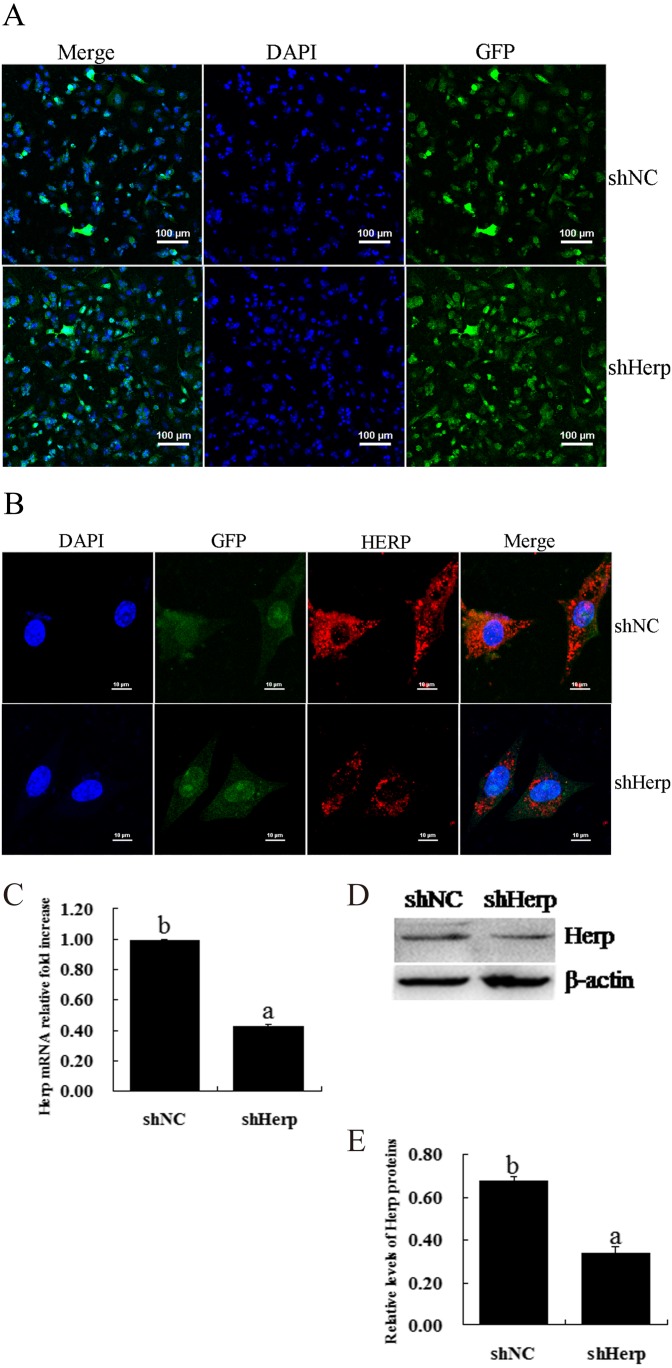
Fig. 2.
Effective inhibition of Herp expression in primary mouse granulosa cells via transduction of the shHerp lentiviruses. A: Fluorescence images of granulosa cells transduced by lentivirus for 48 h. Scale bars, 100 μm. B: Immunofluorescent analysis of Herp protein expression levels in granulosa cells transduced with the shHerp lentivirus for 48 h. C: Relative mRNA expression of the Herp gene in granulosa cells transduced with the shHerp lentivirus for 48 h. The amounts of mRNA were normalized to that of β-actin. D and E: Western blot analysis of Herp protein expression levels in granulosa cells transduced with shHerp lentiviruses for 48 h. The statistical analysis is shown in the bar graphs. Data are presented as the mean ± SEM. Bars with different letters are significantly different (P < 0.05).

