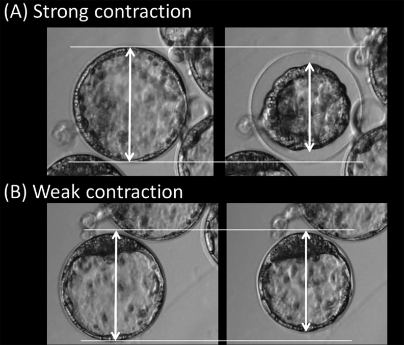
Fig. 2.
Definition of contraction types. All images were taken at the same magnification and in the same field of view. The images show contraction from the anteroposterior view of the blastocyst. Arrows indicate the diameter of each blastocyst. (A) An example of strong contraction: the volume reduction is greater than 20%, and is associated with expansion of the perivitelline space. This example shows a 41% volume reduction. (B) Weak contraction: the volume reduction is less than 20%, and is not associated with expansion of the perivitelline space. This example shows a 17% volume reduction.

