Figure 4.
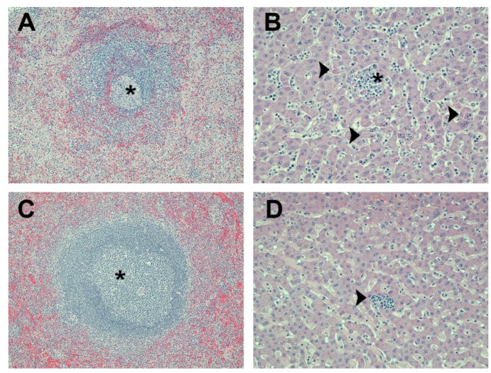
Photomicrographs of liver and spleen from macaques that were challenged with MARV. (A) spleen from a control animal (vaccinated with QS-21 adjuvant only and challenged subcutaneously) has a lymphoid nodule with markedly reduced numbers of lymphocytes (i.e., lymphoid depletion) and which is surrounded by pale pink deposits of fibrin in the adjacent red pulp. The center of this nodule (asterisk) has an aggregate of histiocytes and fragments of lysed lymphocytes; (B) liver from a control animal (vaccinated with QS-21 adjuvant only and aerosol challenged) has multiple foci of hepatocellular degeneration and necrosis (arrowheads). Numerous neutrophils and monocytes are present in the sinusoidal blood vessels and aggregate around necrotic hepatocytes in one area (asterisk); (C) spleen from a VLP-vaccinated animal (Poly I:C adjuvant and aerosol challenged) that survived the viral challenge. A proliferation of lymphocytes (i.e., lymphoid hyperplasia) expands a lymphoid nodule (asterisk); (D) liver from a VLP-vaccinated animal (QS-21 adjuvant and aerosol challenged) that survived the viral challenge. Tissue is normal except for a small focus of subacute inflammation (arrowhead). Magnification: 100× for panels (A) and (C); 200× for panels (B) and (D). Hematoxylin and eosin stain was used for tissue sections in all panels.

