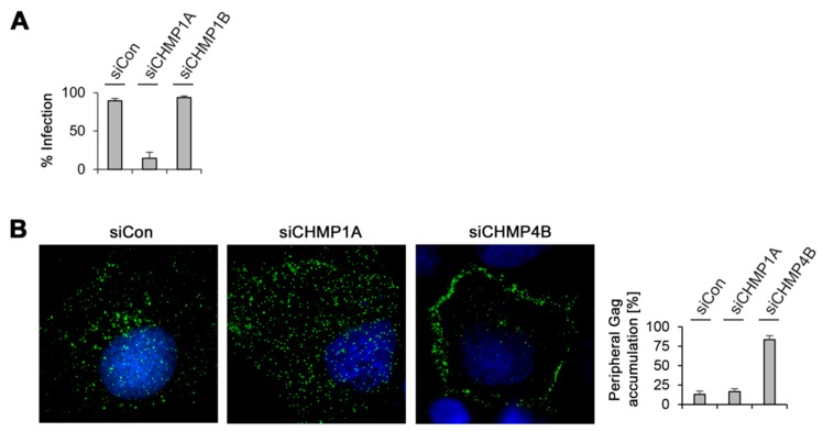
Figure 7.
MLV infectivity and intracellular distribution in CHMP-knockdown cells. (A) Depletion of CHMP1A, but not of CHMP1B, decreased MLV infectivity. Control-, CHMP1A-, and CHMP1B-knockdown HuH-7 cells were cotransfected with MLV.WTFLAG/HA plus a pCL-MFG-LacZ reporter plasmid. Viral particles released into the supernatants were harvested 24 h after DNA transfection and transduced into murine NIH 3T3 cells. Cells were stained for ß-galactosidase activity two days later, quantitated, and averaged from two independent experiments; (B) Control-, CHMP1A-, and CHMP4B-knockdown HuH-7 cells were transfected with MLV.WTFLAG/HA and subjected to immunostaining with mouse anti-FLAG antibodies. Following staining with AlexaFluor 488-conjugated anti-mouse antibodies, cells were analyzed by fluorescence microscopy and representative images are shown. DNA staining is shown in blue. For quantification, the Gag distribution pattern was inspected in about 50 cells from two independent experiments and plotted in the graph.

