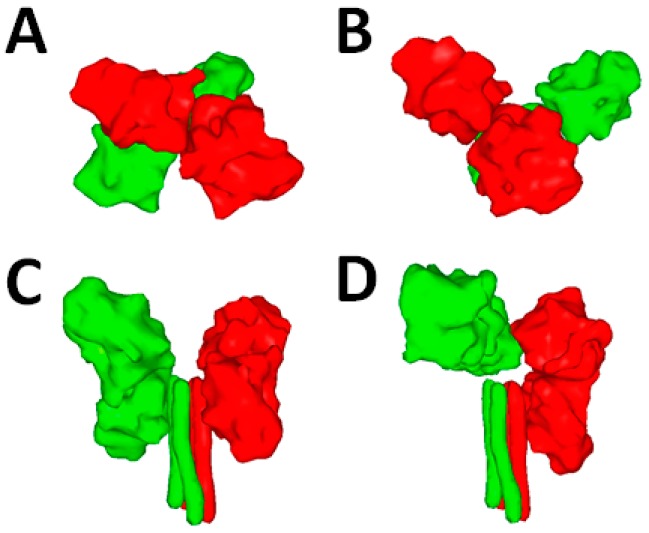
Figure 2.
Possible structure(s) of the MeV attachment protein. (A and B) Crystal structures of MeV H in complex with the receptor signaling lymphocyte activation molecule (SLAM) (not shown). Two tetrameric configurations were determined (PDB 3ALZ and 3ALX). High-resolution structures were morphed into low-resolution images using the Sculptor software package; (C and D) Two alternative folds of H tetramers. Images were obtained from homology models performed with the canine distemper virus CDV) H sequence (strain A75/17) and based on the atomic coordinates of Newcastle disease virus (NDV) HN (PDB 3T1E) and hPIV5 HN (PDB 4JF7). The dimers of the attachment protein tetramer are color-coded in green and red.

