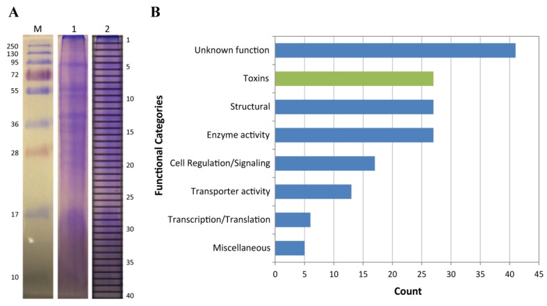
Figure 3.
Venom proteome of C. fuscescens. (A) SDS-PAGE analysis of crude venom (lanes 1 and 2). The 40 gel bands used for in-gel tryptic digestion and tandem mass spectrometry are indicated in lane 2. Molecular masses of the protein marker (M) are shown alongside in kDa; (B) Functional annotation of proteins identified in proteomics experiments.

