
Figure 4. Microinjection of CRISPR-Cas9 reagents into the mouse zygote.
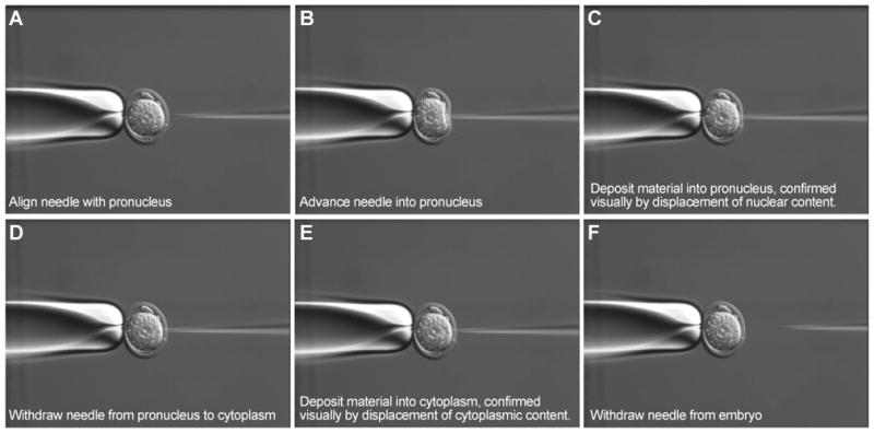
Panels “A” to “F” show in frames how to deposit the CRISPR-Cas9 reagents into both the pronucleus and the cytoplasm.
Official websites use .gov
A
.gov website belongs to an official
government organization in the United States.
Secure .gov websites use HTTPS
A lock (
) or https:// means you've safely
connected to the .gov website. Share sensitive
information only on official, secure websites.
Panels “A” to “F” show in frames how to deposit the CRISPR-Cas9 reagents into both the pronucleus and the cytoplasm.