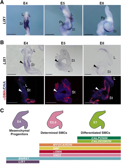
Fig. 1.

Transient expression pattern of LIX1 in the developing chick stomach. a LIX1 whole-mount in situ hybridization of embryonic day 4 (E4) to E6 stomachs. Scale bars, 1 mm. b Serial longitudinal sections of E4 to E6 stomachs analysed by in situ hybridization using the LIX1 riboprobe and by immunofluorescence with anti-αSMA antibodies. Nuclei are visualized with Hoechst. Black arrowheads show the mesenchymal expression of LIX1 at these stages. White arrowheads show the absence of αSMA in the LIX1-expressing domains. Scale bars, 500 μm. c Cartoon illustrating the steps of stomach mesenchyme development and summarizing Fig. 1a, b and Additional file 1: Figure S1A. L, Lung; St, Stomach; Pyl, Pylorus
