Fig. 4.

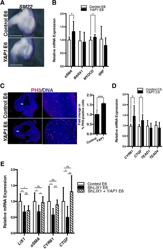
YAP1 stimulates stomach mesenchymal progenitor proliferation and smooth muscle cell determination. a Whole-mount in situ hybridization of E6 stomachs using the SM22 riboprobe. White dashed lines indicate the position of the intermuscular tendon in the stomach. Scale bars, 1 mm. b RT-qPCR analysis of relative mRNA expression in E6 stomachs. Data were normalized to GAPDH expression. Normalized expression levels were converted to fold changes. Values are presented as the mean ± standard derivation (SD) of n = 8 controls vs. n = 6 YAP1-expressing stomachs. *P < 0.05 by one-tailed Mann–Whitney tests. c Serial transverse sections of E6 stomachs analysed by immunofluorescence using anti-PH3 antibodies. Nuclei are visualized with Hoechst. White arrows in the left panels indicate the area imaged at high power in right panels. Graph represents the quantification of PH3-positive cells. Normalized expression levels were converted to fold changes. Values are presented as the mean ± standard error of the mean of n = 10 controls vs. n = 10 YAP1-expressing stomachs. ****P < 0.0001 by two-tailed Mann–Whitney test. d RT-qPCR analysis of relative mRNA expression in E6 stomachs. Data were normalized to GAPDH expression. Normalized expression levels were converted to fold changes. Values are presented as the mean ± SD of n = 8 controls vs. n = 6 YAP1-expressing stomachs. *P < 0.05 by one-tailed Mann–Whitney test. e RT-qPCR analysis of relative mRNA expression in E6 stomachs. Data were normalized to GAPDH expression. Normalized expression levels were converted to fold changes. Values are presented as the mean ± SD of n = 8 controls vs. n = 8 ShLIX1 vs. n = 6 ShLIX1 + YAP1-expressing stomachs. *P < 0.05; **P < 0.01 by one-tailed Mann–Whitney test. ns, not significant
