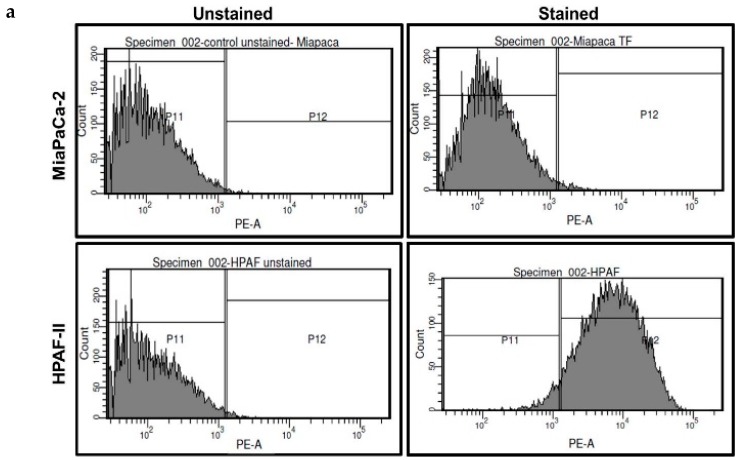
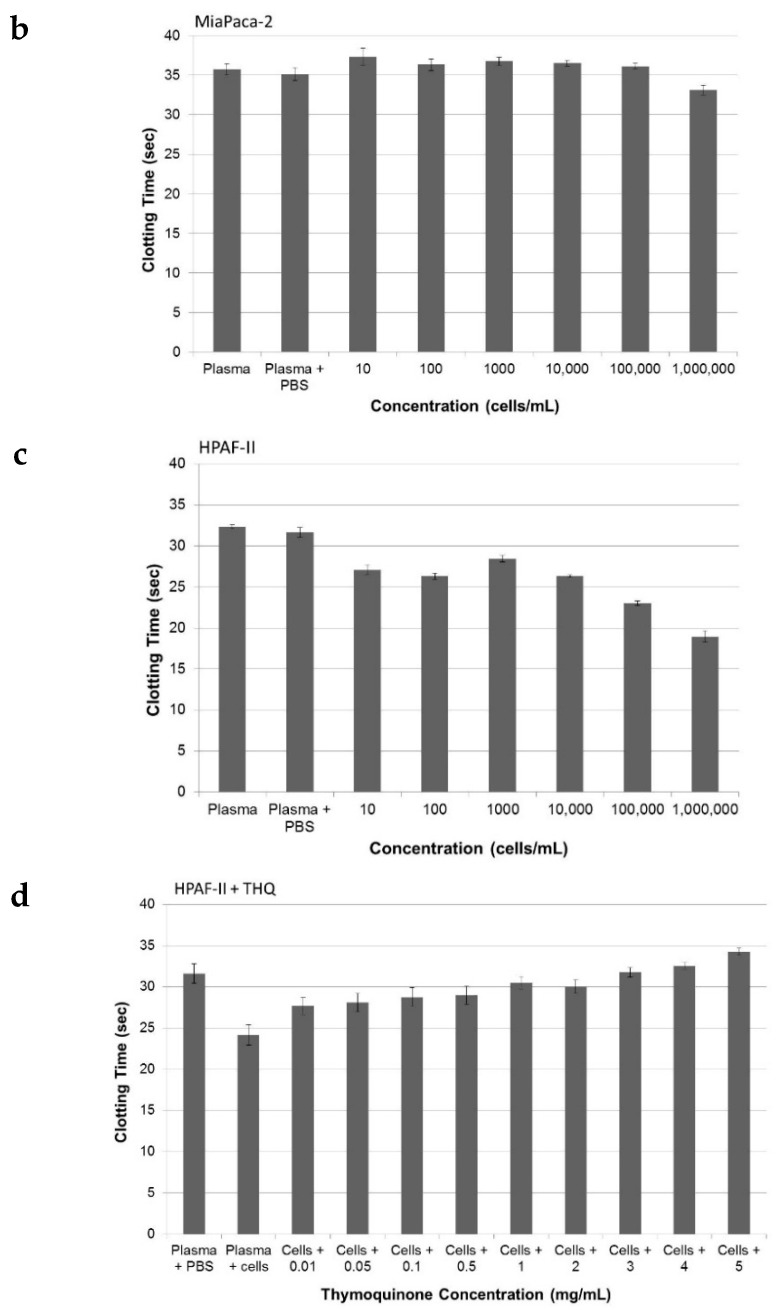
Figure 3.
THQ reverses coagulation mediated by tissue factor (TF) present on pancreatic cancer cells. (a) Flow cytometry analysis of TF on pancreatic cancer cells. Data in the top panels are from MiaPaCa-2 cells, and data in the bottom panels are from HPAF-II cells. The left panels serve as a control from cells unstained for TF, and the right panels are from cells stained with TF-antibody, followed by PE-conjugated secondary antibody. The top right panel shows the absence of TF on MiaPaCa-2 cells. Presence of TF on HPAF-II cells is confirmed by the TF staining on HPAF-II cells (bottom, right panel). Approximately 98% of HPAF-II cells were positive for the presence of TF; (b) MiaPaCa-2 cells do not cause coagulation of plasma. The coagulation time measured with aPTT assay did not show any change in the presence of an increasing number of MiaPaCa-2 cells; (c) the coagulation time measured with aPTT assay showed that HPAF-II cells can decrease the coagulation time. Both t-test, as well as ANOVA with post hoc (Tukey), showed that there was no significant difference in the clotting time between the plasma and plasma with PBS. Addition of cells (10 to 10,000) in each case significantly (p < 0.001) reduced the clotting time compared to PBS-plasma; (d) THQ reversed the coagulation induced by HPAF-II cells. To 1 × 10⁶ HPAF-II cells, THQ was added in increasing concentration, from 0.01 mg/mL to 5 mg/mL. Both t-tests, as well as ANOVA with post hoc (Tukey), showed that THQ at each concentration (0.01 to 5 mg/mL) significantly (p < 0.001) reduced the clotting time compared to plasma + cells with no THQ. The experiments were performed three times in triplicate. Error bars show the standard deviations of the mean values.


