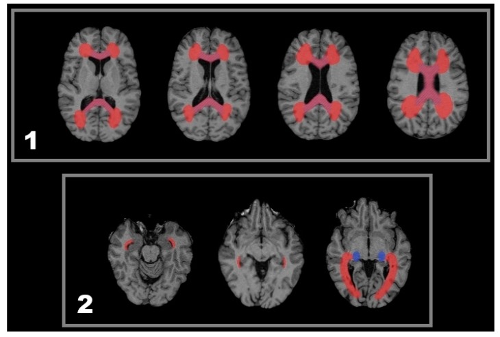
Figure 5.
Sketch of anatomical regions of interest regarding the statistical analysis of lesion-related volume decrease. (1) The red coloured region delineates a WM region that contains callosal fibres including an adjacent delta-like extension and was searched for lesions. The region investigated for callosal atrophy is drafted in blue hachures; (2) In these exemplary slices, a red draft delineates the region covering all three bundles of the optic radiation including the entire lateral and superior wall of the inferior horn of the lateral ventricles [36,37,38,39,40]. For the LGN, a high inter-individual variability is known regarding both size and location [37]. Therefore, a region of the posterior thalamus containing the LGN derived from a population map by Bürgel et al. was approximated for each patient so as to assess LGN atrophy consistently (blue draft).

