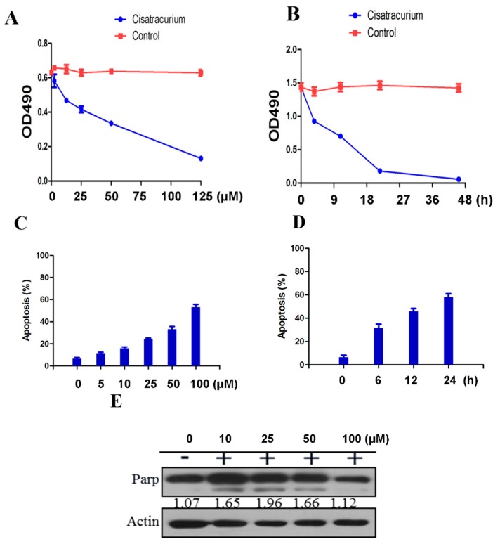
Figure 3.
Cisatracurium causes inhibition of cell proliferation and induction of cell death. (A) HUVEC was incubated with cisatracurium (0, 5, 10, 25, 50, or 125 µM) for 12 h. Cell viability was analyzed by CCK assay; (B) HUVEC was incubated with cisatracurium (10 µM) for 0, 6, 12, 24, or 45 h. Cell viability was analyzed by CCK assay; (C) HUVEC was incubated with cisatracurium (0, 5, 10, 25, 50, or 100 µM) for 12 h. Cells were stained by PI and Annexin-5 and then analyzed by flow cytometry to calculate the rate of apoptosis. Data are presented as mean ± SD from three independent experiments. p < 0.01; (D) HUVEC was incubated with cisatracurium (5 µM) for 0, 6, 12, or 24 h. Cells were stained with PI and Annexin-5, then analyzed by flow cytometry to calculate the rate of apoptosis. Data are presented as mean ± SD from three independent experiments. p < 0.01; (E) HUVEC was incubated with cisatracurium (0, 10, 25, 50, 100 µM) for 12 h. Samples were then subjected to immunoblot using antibodies against PARP or β-actin. Numbers indicate the normalized optical density ratio of cleaved PARP to β-actin. Data are presented as a representative image from three independent experiments.

