Figure 4.
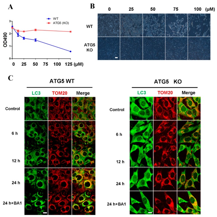
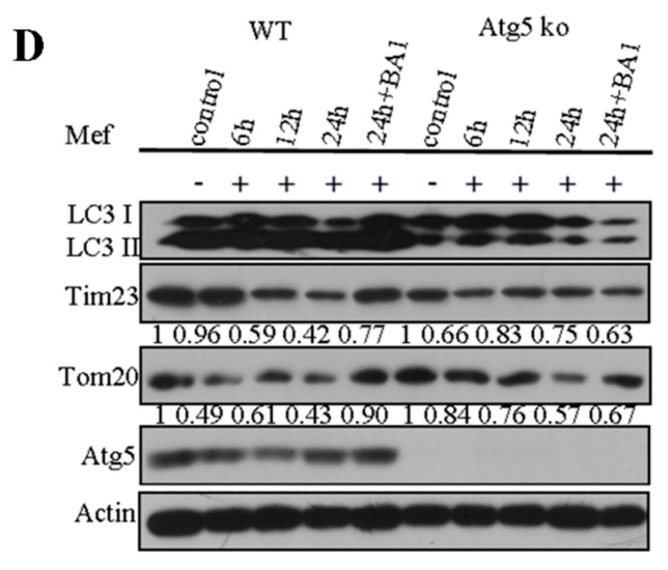
Inhibition of cell proliferation and induction of cell death by cisatracurium is blocked in ATG5-deficient cells. (A) WT or ATG5 KO MEFs were incubated with cisatracurium (0, 10, 25, 50, or 125 µM) for 12 h. Cell viability was analyzed by CCK assay; (B) WT or ATG5 KO MEFs were incubated with cisatracurium (0, 25, 50, 75, or 100 µM) for 12 h. Cells were observed by light microscopy. Bar = 100 µm; (C) WT or ATG5 KO MEFs were incubated with cisatracurium (5 µM) for 0, 6, 12, or 24 h in the presence or absence of Baf A1. Cells were stained by anti-LC3 (green) and anti-TOM20 (red) antibody. Bar = 15 µm; (D) WT or ATG5 KO MEFs were incubated with cisatracurium (5 µM) for 0, 6, 12, or 24 h in the presence or absence of Baf A1. Samples were then subjected to immunoblot using antibodies against LC3, Tim23, TOM20, ATG5 or β-actin. Numbers represent the normalized optical density ratio of indicated bands to β-actin. Data are presented as a representative image from three independent experiments, and the intensity of indicated bands was measured with ImageJ software and normalized by Beta-actin.


