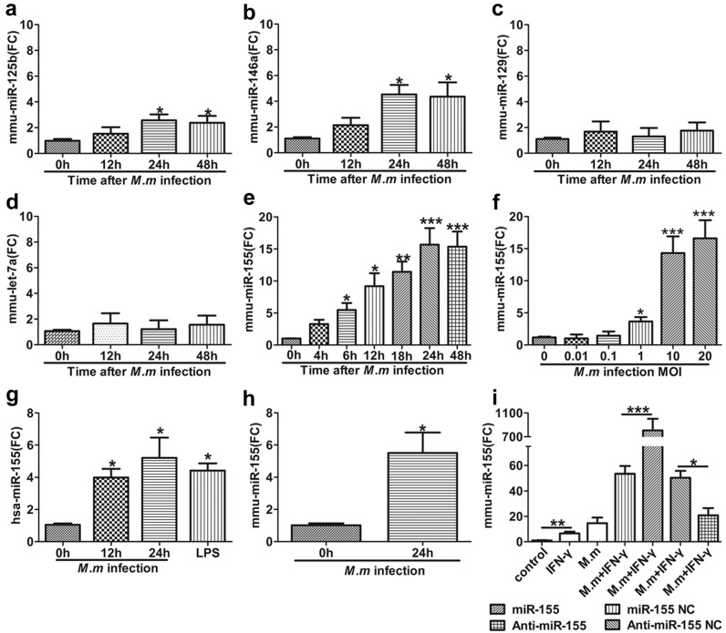
Figure 1.
Distinguishing expression of miRNAs in M.m infected macrophages. (a–e) Expression of miR-125b, miR-146a, miR-129, Let-7a and miR-155 in RAW 264.7 cells after M.m infection for the indicated times; (f) Expression of miR-155 in RAW 264.7 cells at different multiplicities of infection (MOI) for 24 h; (g) miR-155 expression levels were detected in THP-1 cells after M.m infection or lipopolysaccharide (LPS) (100 ng/mL) treatment; (h) miR-155 expression level was examined in mouse peritoneal macrophages (MPMs); (i) miR-155 expression was detected in RAW 264.7 cells after transfected with miR-155 Negative Control (NC), miR-155, anti-miR-155 NC and anti-miR-155 prior to infection with M.m or treatment with IFN-γ/M.m for 24 h. All miRNAs were detected by quantitative real-time polymerase chain reaction (PCRs) (qPCRs). U6 snRNA was used as endogenous control. Experiments were executed with at least three biologically independent replicates, data were shown as the mean ± SEM. * p < 0.05, ** p < 0.01, *** p < 0.001. FC: fold change.

