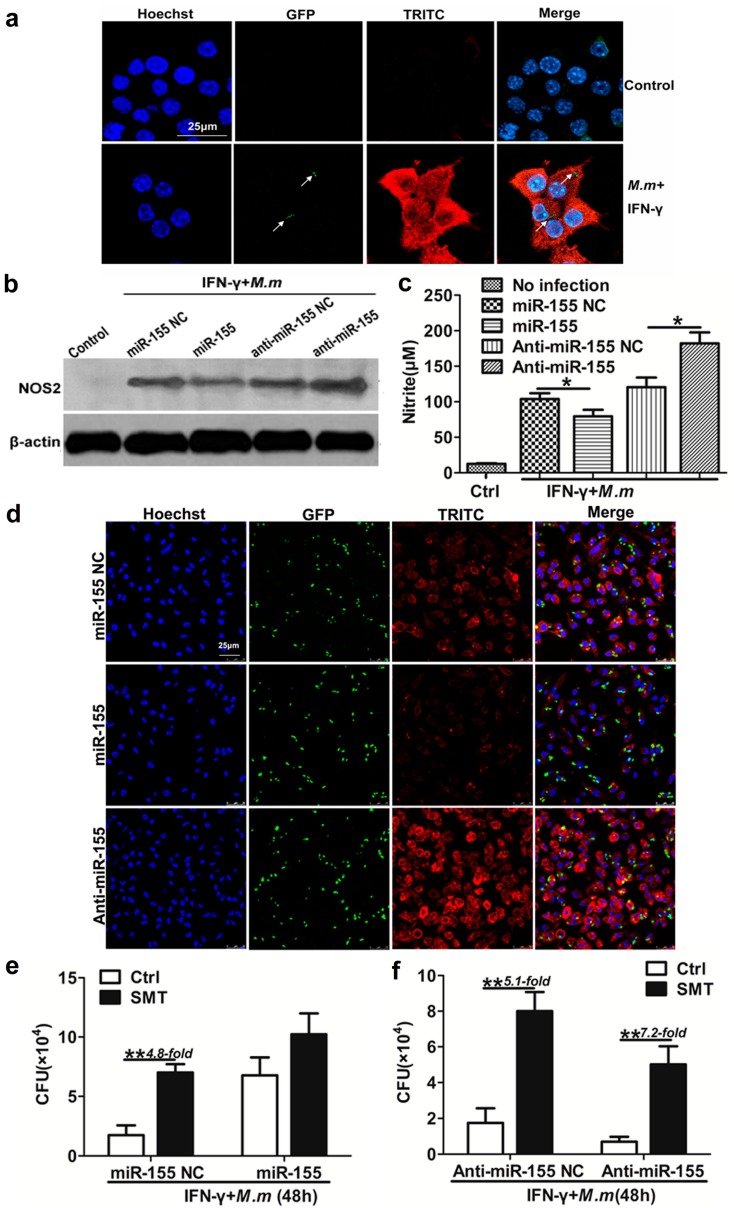
Figure 3.
miR-155 promote M.m survival in macrophages by inhibition of NO. (a) NOS2 expression in the cytoplasm of RAW 264.7 cells. The co-localization of GFP-labeled M.m (green) and TRITC-labeled NOS2 (red) in the cytoplasm was examined by confocal microscopy. Arrows indicate M.m. Nucleus of RAW 264.7 cells were stained with Hoechst (purple); (b) Immunoblot blot analysis of NOS2 in RAW 264.7 cells; (c) NO synthesis in RAW 264.7 cell. Cells were transfected with miR-155 NC, miR-155, anti-miR-155 NC and anti-miR-155 followed by IFN-γ treatment and infected with M.m for 24 h, supernatant nitrite was determined by the Griess test; (d) NOS2 expression in the cytoplasm of MPM cells. Cells were transfected with miR-155 NC, miR-155, anti-miR-155, and infected with GFP-labeled M.m/IFN-γ for 24 h. The co-localization of M.m (green) and TRITC-labeled NOS2 (red) in the cytoplasm were detected by confocal microscopy. Nucleuses of MPMs were stained with Hoechst (purple); (e,f) The effect of miR-155, anti-miR-155 and NOS2 inhibitor on M.m survival in RAW 264.7 cells. Cells were transfected with miR-155, anti-miR-155 and corresponding NC, pretreated with 1 mM SMT for 2 h, infected with IFN-γ/M.m for 48 h. Intracellular M.m survival was determined by CFU assay. Data are shown as the means ± SEM of three independent experiments, * p < 0.05, ** p < 0.01.

