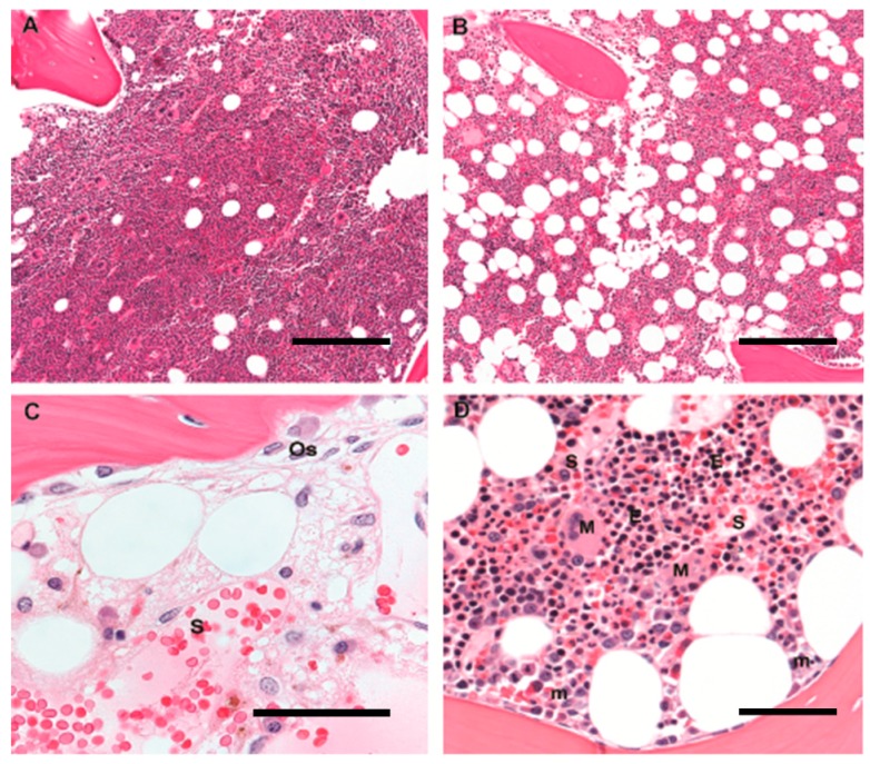
Figure 1.
Human marrow architecture in youth and age: Hematopoiesis in human adults is predominantly axial, and diagnostic bone marrow biopsies sample the pelvic iliac crest. Trilineage hematopoiesis is admixed with increasing amounts of mature adipose tissue with age; adipocytes appear as round clear spaces. (A) A bone marrow biopsy from a 5-year old child is >90% cellular, with a predominance of trilineage hematopoiesis and little admixed adipose tissue; original magnification 10×; scale bar 100 μm; (B) A bone marrow biopsy from a 60-year old adult is composed of 50% hematopoietic elements and 50% admixed mature adipose tissue; original magnification 10×; scale bar 100 μm; (C) A post-chemotherapy-marrow reveals the underlying bone marrow architecture and microenvironment. Trabecular bone is curvilinear lamellar bone with apposed osteoblasts (Os), and a thin osteoid seam of unmineralized collagen. Dilated thin-walled sinusoids (S) are filled with red blood cells and have closely-apposed stromal cells with ovoid nuclei. Scattered mononuclear cells include plasma cells, mast cells, and macrophages, some of which contain yellow-brown hemosiderin pigment; original magnification 60×; scale bar 25 μm; (D) Erythroid colonies (E) appear as colonies of round cells with dark nuclei and are located away from trabecular bone, close to thin-walled sinusoidal vessels (S); megakaryocytes (M) are likewise located in close contact with sinusoids whereas immature myeloid precursors (m) are localized near trabecular bone. Original magnification 20×; scale bar 50 μm.

