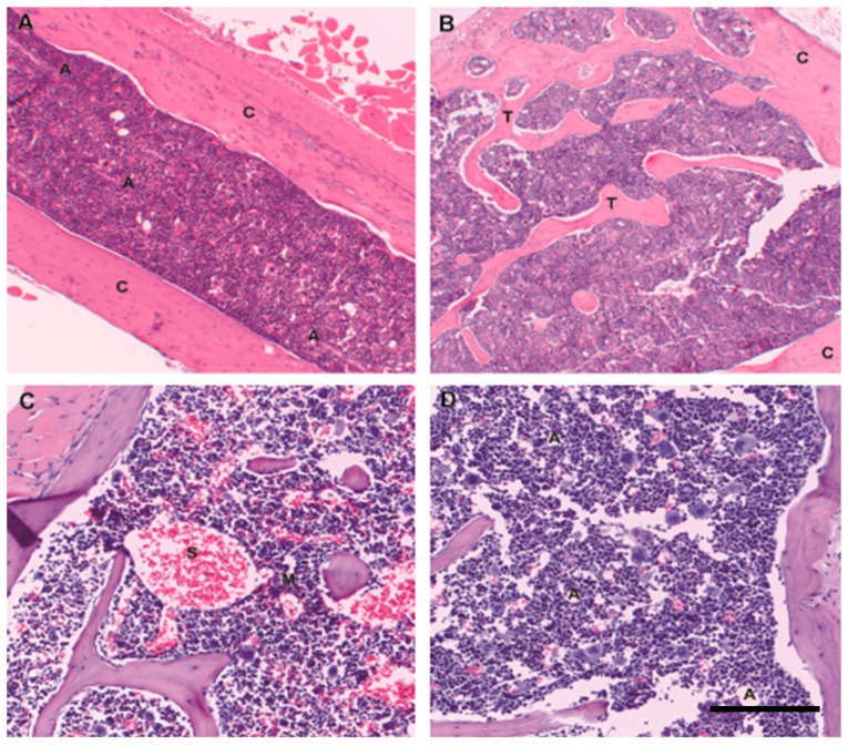
Figure 2.
Mouse marrow architecture in youth and age: Hematopoiesis in mice is present throughout the skeleton. Photomicrographs from C57BL6 wild-type animals. (A) Mouse hematopoietic marrow architecture is most commonly evaluated in whole mounts of tubular bone (femur), where predominantly cortical bone (C) in contact with hematopoietic marrow. Small-diameter arterioles (A) with well-defined multilayered walls run the length of the femur; (B) Flat bone sites such as pelvis in contrast more closely resemble the trabecular-bone (T) predominant architecture of adult human hematopoietic marrow; cortical bone, (C); (C) Mouse sternum also has a significant trabecular component; in 8 week-old mice hematopoietic cellularity is close to 100%, with no identifiable admixed fat. Similar to human marrow megakaryocytes (M) are closely opposed to thin-walled and sometimes ectatic sinusoidal vessels (S); (D) Similar to human marrow, mouse marrow increases in fat content with age, although the absolute percentage of mature adipose tissue is much lower than that seen in older adult humans. Here a two-year old mouse shows scattered admixed adipocytes (A), accounting for 5%–10% of marrow cellularity. Original magnification 4×; scale bar 200 μm.

