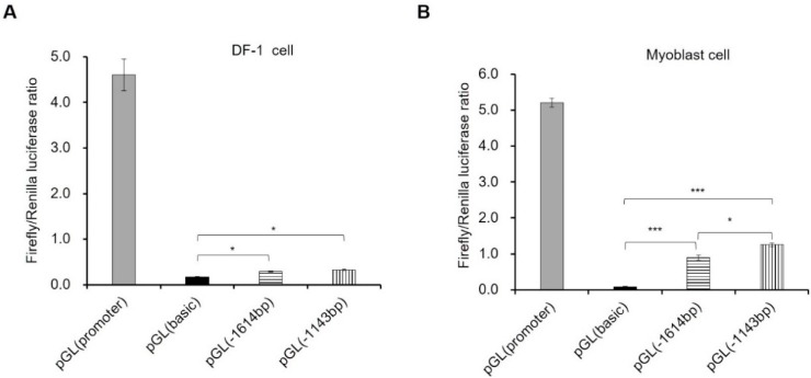
Figure 1.
Characterization of promoter activity of gga-miR-206 in DF-1 and myoblast cell. (A) Detection of miR-206 promoter activity in DF-1 cell lines; (B) Detection of miR-206 promoter activity in myoblast cells. Myoblast cell was isolated from skeletal muscle of 11-day-old embryos. The ratio of firefly and renilla luciferase was used for detecting the promoter activity through co-transfecting pGL3.0 and pGL-TK (as normalize control). In this panel, data are presented as mean ± standard error (SE). * p < 0.05 and *** p < 0.001 were estimated by Student’s t-test (n = 3).

