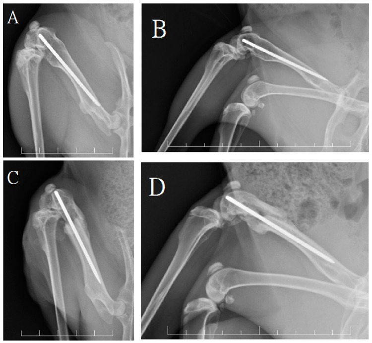
Figure 3.
The radiographic assessments at 24 weeks. The anterioposterior (A) and lateral (B) view of a rabbit in group A showed good bone growth within the bone defect site with intact anterior cortical integrity; The anterioposterior (C) and lateral (D) view of a rabbit in group B showed callus formation but had broken of the anterior cortex. Each frame rates one centimeter.

