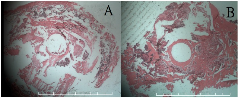
Figure 4.
Histological results (200×) of coronal section on femoral bone defect site with hematoxylin and eosion (H & A) stain. (A) The results at 12 weeks showed no inflammatory overreaction around the inferior aspect of the defect site; (B) The results at 24 weeks revealed good bone callus formation around the entire defect site. Each frame rates one millimeter.

