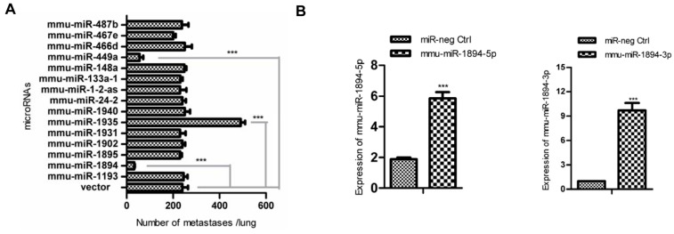
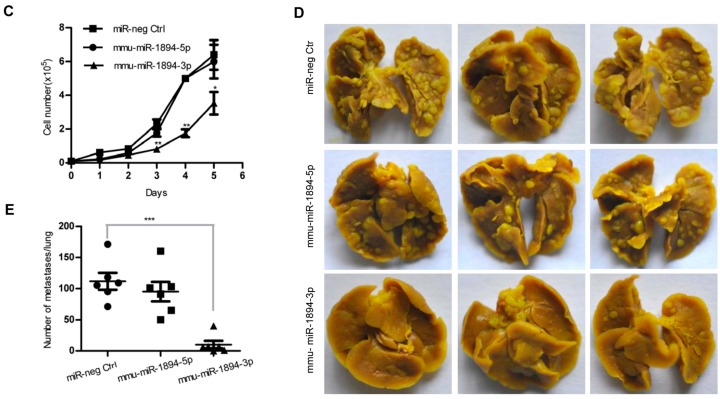
Figure 1.
Mouse microRNA screening and lung metastasis assay. (A) The numbers of metastasis nodules in lungs were calculated 14 days after transplantation with the indicated microRNA or empty vector in 4TO7 cell lines (n = 6 for each group). *** indicates p < 0.001 versus vector control; (B) The expression of mmu-miR-1894-3p and mmu-miR-1894-5p in 4TO7 stable cell lines was determined by qRT-PCR. U6 snRNA was used for normalization. *** indicates p < 0.001 versus control; (C) Growth curves of the 4TO7 stable cell lines expression of mmu-miR-1894-3p and mmu-miR-1894-5p at indicated time. * indicates p < 0.05, ** indicates p < 0.01 versus control; (D) Representative photos for lung metastasis nodules. 4TO7 cells expressing mmu-miR-1894-3p or mmu-miR-1894-5p were injected into the tail veins of Balb/c female mice. Two weeks later, the lungs were fixed in Bouin’s solution and photographed; and (E) The numbers of metastasis nodules in (D) were calculated (n = 6 for each group). *** indicates p < 0.001 versus control.


