Figure 4.
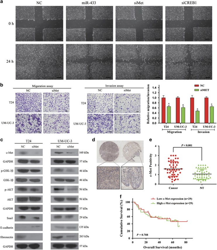
Knockdown of c-Met suppresses BCa cells motility. (a) UM-UC-3 cells were transfected with NC, miR-433 mimics, siMet, and siCREB1, respectively. Wound-healing assay was performed with a 24-h recovery period. (b) Transwell assay (representative micrographs were presented). siMet impaired the motility of T24 and UM-UC-3 cells. (c) siMet inhibited EMT and AKT/GSK-3β signaling-related proteins in T24 and UM-UC-3 cells. (d) Representative images of IHC staining of TMA. c-Met showed a membranous and cytoplasmic location. (e) Statistical analysis indicated that the expression level of c-Met protein in BCa tissues was significantly higher than that in adjacent non-tumor tissues. (f) Kaplan–Meier survival analysis. The protein expression of c-Met was not associated with the overall survival rate in BCa patients. Error bars represent the S.E. obtained from three independent experiments; *P<0.05. Scale bars=100 μm.

