Figure 8.
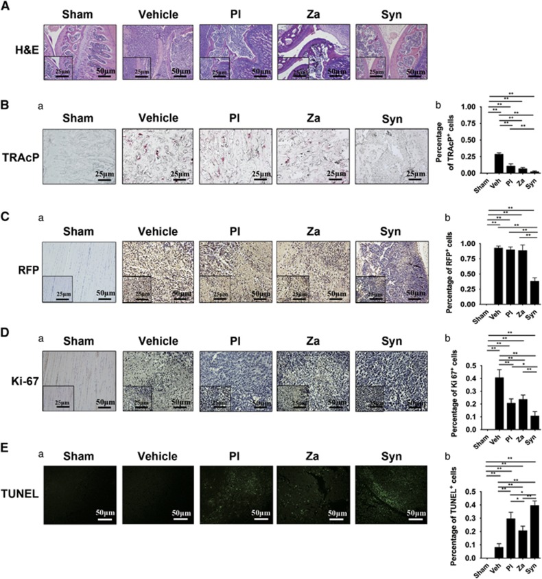
In vivo potentiated antiosteoclastogenesis and antitumorigenesis through combined treatment with PL and ZA. (A) H&E staining of tibia in tumor-bearing mice after 6 weeks of treatment (magnification, × 200, scale bar=50 μm; magnification, × 400, scale bar=25 μm) (Ba) TRAcP-positive osteoclasts in the tibiae of tumor-bearing mice after 6 weeks of treatment (magnification, × 400, scale bar=25 μm). (b) Histomorphometric quantifications were calculated. (Ca) Immunohistochemistry for RFP in tumor tissues (magnification, × 200, scale bar=50 μm; magnification, × 400, scale bar=25 μm). (b) Histomorphometric quantifications were calculated. (Da) Immunostaining of Ki-67-positive cells in tumor tissues (magnification, × 200, scale bar=50 μm; magnification, × 400, scale bar=25 μm). (b) Histomorphometric quantifications were calculated. (Ea) Immunohistochemistry indicating TUNEL-positive cells in tumor tissues (magnification, × 200, scale bar=50 μm). (b) Histomorphometric quantifications were calculated. The data are presented as the means±S.D. Each group contained 10 animals (*significant difference at P<0.1; **significant difference at P<0.05)

